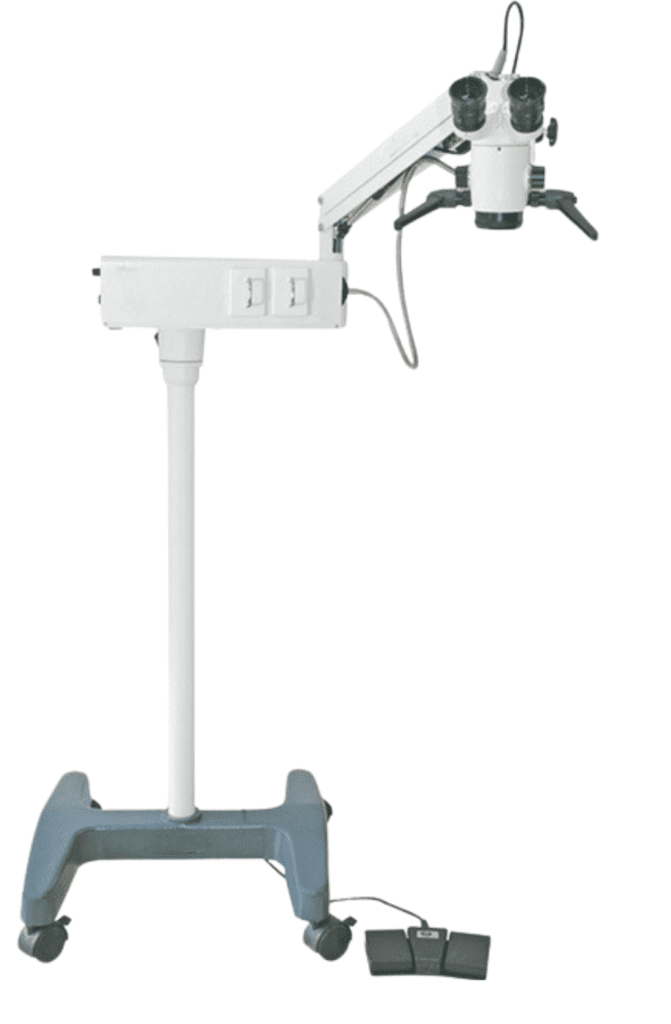
OPHTHALMIC OPERATING MICROSCOPES
An ophthalmic surgical microscope is an instrument that combines lenses and allows an ophthalmologist a stereoscopic(3D), magnified, high-quality image of the small structures around and within the eye during eye surgeries enabling precise manipulation of tiny and intricate structures like the cornea, lens, retina, or optic nerve
We offer a range of state-of-the-art ophthalmology microscopy solutions that support ophthalmic surgeons in achieving optimal patient outcomes in anterior and posterior segment surgery.
Our ophthalmic microscopes are world-renowned for premium optics and illumination that provide outstanding image quality.
Key Features of an ophthalmic operating microscope:
- Magnification:
Variable levels (typically 5x to 40x), allowing the surgeon to zoom in on tiny eye structures. - Binocular viewing:
Provides a stereoscopic (3D) image for depth perception, crucial for microsurgery. - Illumination:
Built-in coaxial (direct) light source, often using halogen, xenon, or LED, for shadow-free lighting. - Foot controls:
Allow the surgeon to adjust focus, magnification, or position without using their hands. - Motorized movement:
Some models have programmable positioning and stabilization. - Assistant viewing systems / Cameras:
Many systems include ports for assistant viewing, teaching, or recording surgeries.
Used in Procedures Like:
- Cataract surgery
- Retinal surgery (e.g., vitrectomy)
- Glaucoma surgery
- Corneal transplantation
- Oculoplastic procedures
Caring for the operating microscope:
- Keep the microscope in a dry, cool and well-ventilated place to prevent fungus growth on the optics (lenses).
- To protect it from dust when not in use, drape a cover over the microscope.
- Wipe down the external surfaces with a lint free cloth.
- Cover the foot pedal with a clear plastic bag to prevent surgical and cleaning fluids from entering and damaging the electronics.
- Use a voltage stabiliser with the microscope. This will prevent sudden increases in voltage from destroying the bulbs and will ensure that the illumination provided remains constant.
- Before using, test the controls of the foot pedal (the x,y movement, zoom, focus, light on and off).
- Before using, check that the suspension arm can be fixed into position to ensure that it does not fall on the patient.
- Avoid kinking or bending the fibre optic cables.
- Do not move the microscope while the bulb is still hot because strong vibrations may damage the filament.
Every six months, clean and oil the wheels and the brakes. Remove any.